Treatment rooms
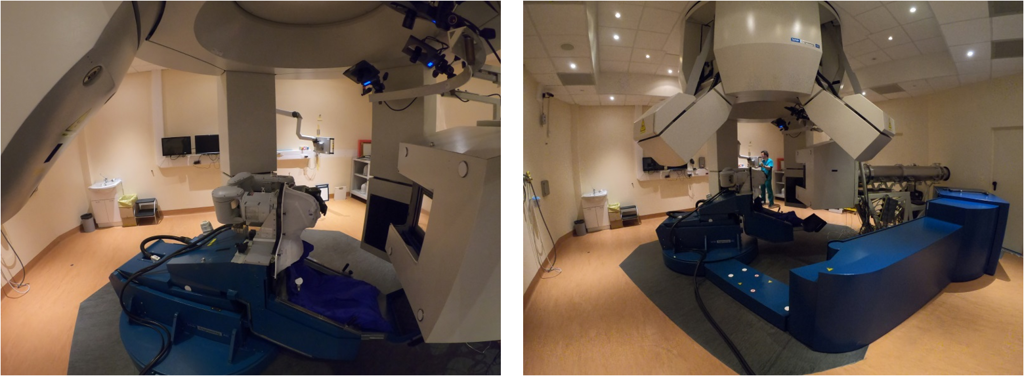
Where the hadrontherapy sessions take place
Patient's procedure
Once the possible enrolment for the treatment has been verified, the simulation phase is carried out after the first medical visit.
The immobilization devices to deliver the treatment are chosen at this stage.
Immobilization devices mean personalised thermoplastic masks and pillows that will keep each patient at the same position all the time; these procedures are not painful at all.
While the patients are wearing these masks, CT and MRI images are taken and used by doctors and physicists to set the best way to irradiate the lesion.
After the simulation stage, the patient goes to the treatment rooms for the therapy.
Therapy can follow different pathways based on the location of the tumour.
Head and neck cancers
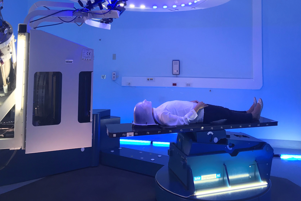
The treatment session begins with the positioning of the patient on the carbon fibre bed which moves by means of a robotic arm (Patient Positioning System or PPS).

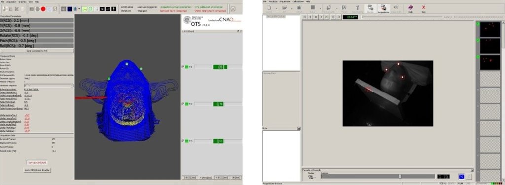
The patient is then immobilized with the mask created during the simulation phase and which will be used for the whole treatment. Optoelectronic markers are stuck to mask and recognized by 3 infrared cameras (Optical Tracking System or OTS), which are necessary to check the patient's position before and during treatment.

Subsequently, two X-ray images are taken through the Patient Verification System or PVS, which will be compared with the images reconstructed by the simulation CT scan. This comparison will result in the displacement to be applied to bring the patient to the position in which the treatment plan was calculated (accuracy 0.3 mm).

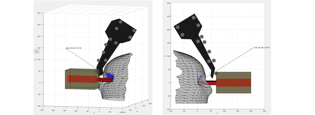
Once the correct positioning has been ascertained, the irradiation begins. The treatment plan subdivides the lesion in various slices. The particle beam irradiates one slice after the another, following the commands determined by the treatment plan. The irradiation of the lesion occurs in certain positions of the PPS that identify the entry of the beam, called fields. Moving from one field to another is carried out by means of the PPS which extrapolates the position from the treatment plan. The irradiation lasts about 3 minutes per field, while the passage between fields takes about 2-3 minutes for each movement. The treatment continues in a number of sessions always determined by the treatment plan studied beforehand.
Pelvic and abdominal cancers
The patient is positioned on the carbon fibre bed through lasers with landmarks performed during the simulation phase. This positioning is carried out in a room adjacent to the treatment room, called the CAPH (Computer Aided Positioning in Hadrontherapy) room.

The patient is then carried with the bed to the treatment room, where the bed is hooked to the robotic arm or PPS (Patient Positioning System).
Similarly to head and neck tumours, two X-ray images are taken for pelvic and abdominal tumours too through the Patient Verification System or PVS, which will be compared with the images reconstructed by the simulation CT scan. This comparison will result in the displacement to be applied to bring the patient to the position in which the treatment plan was calculated (accuracy 0.3 mm).
For the pelvic and abdominal area, an internal organs control is also performed given their possible motility through volumetric images, always acquired with the PVS. Once the correct positioning has been ascertained, the irradiation begins as described for head tumours.

Eye cancer
Two simulation CTs are performed to define the optimal direction of the gaze and at that moment the thermoplastic mask is made to carry out the treatment. The patient is not immobilized on the carbon fibre bed, but uses a chair-shaped structure, stuck to the robotic arm (Patient Positioning System or PPS).
Once the mask is put on, the treatment involves a series of controls that require good collaboration from the patient, given the particular position of the lesion. Positioning control is carried out with the acquisition of X-ray images, through the Patient Verification System (PVS), prior to treatment and during irradiation. This control focuses on checking the position of surgical clips positioned before the simulation phase by the specialist in eye surgery.

During the entire duration of the treatment, the movement of the eye is controlled through the Eye Tracking System (ETS), so as to be able to give the patient commands and verify that the gaze is directed in the correct way.

Treatment plan involves four treatment sessions.


